Digital Pathology Is Moving Upstream: Why the Next Workflow Layer Is Digital Staining
Author
Published

Digital pathology is entering a new phase. For years, the category was mostly associated with slide scanners, image viewers, digital archives, and the shift from glass slides to whole-slide images. This is all still important, without reliable image capture, storage, viewing, and workflow software, there is no digital pathology stack.
However, we are now moving beyond digitization alone.
One example, Roche recently announcing an agreement to acquire PathAI. Roche described PathAI as a digital pathology and AI-powered technology company serving pathology laboratories and the biopharma industry. The announcement highlighted PathAI’s image management system, AI analysis, workflow capabilities, companion diagnostics, biomarker discovery, and biopharma services.
Digital pathology is not just a question of whether tissue slides can be scanned. It is becoming an infrastructure layer for pathology workflows, clinical development, biomarker discovery, and AI-enabled decision support.
At PathScience, we see another layer emerging in parallel: moving digital pathology upstream.
Instead of only asking what AI can do after a slide has been stained and scanned, Direct-to-Digital Staining asks: What if digital tools could help create the stain-like image itself?
The first wave: digitizing the slide
The first wave of digital pathology focused on converting physical pathology slides into digital images.
This is very important work. Digital slide images can support remote review, archiving, collaboration, case management, education, and AI-assisted analysis. Roche describes digital pathology as enabling high-resolution digital images from physical tissue on slides, allowing pathologists to use AI tools to help facilitate diagnostic workflows.
This first wave helped pathology move from a microscope-only environment toward a software-enabled environment.
But in most digital pathology workflows, the image still begins the same way it has for decades: tissue is processed, chemically stained, placed on a slide, and then scanned.
That means much of the laboratory workflow remains tied to the traditional staining process.
The next wave: rethinking how images are created
Once pathology becomes digital, a new set of questions becomes possible.
Can some staining-related steps be reduced, accelerated, or made more consistent?
Can a single tissue section support more than one type of image output?
Can tissue be preserved for additional analysis?
Can pathologists review familiar images while labs reduce operational bottlenecks?
Can digital tools support the creation of the image, not just the review of the image?
This is where digital staining comes in.
Broadly, virtual or digital staining refers to methods that use trained computational models to generate histological stain images digitally. A 2023 review in Light: Science & Applications describes virtual staining as methods that digitally generate histological stains using trained deep neural networks, including both label-free staining and stain-to-stain transformations.
That distinction is important.
Traditional digital pathology usually starts after staining. Digital staining moves upstream, closer to the point where the pathology image is created.
What PathScience means by Direct-to-Digital Staining
PathScience is focused on Direct-to-Digital Staining.
In simple terms, the workflow is designed to capture images of unstained tissue and use AI-enabled image generation to produce H&E-style virtual stain images for pathology review.
The goal is not to replace pathologists, or autonomous diagnosis, and not simply color correction..
The goal is to generate familiar, interpretable, stain-like images while reducing reliance on traditional chemical staining steps.
This is how PathScience is different than image management platforms or post-scan diagnostic algorithms. Those tools are often focused on what happens after the stained slide is scanned. Direct-to-Digital Staining is focused on what happens before and/or during image creation.
Where digital staining fits in the workflow stack
Digital pathology is not one product category. It is becoming a stack of connected layers.
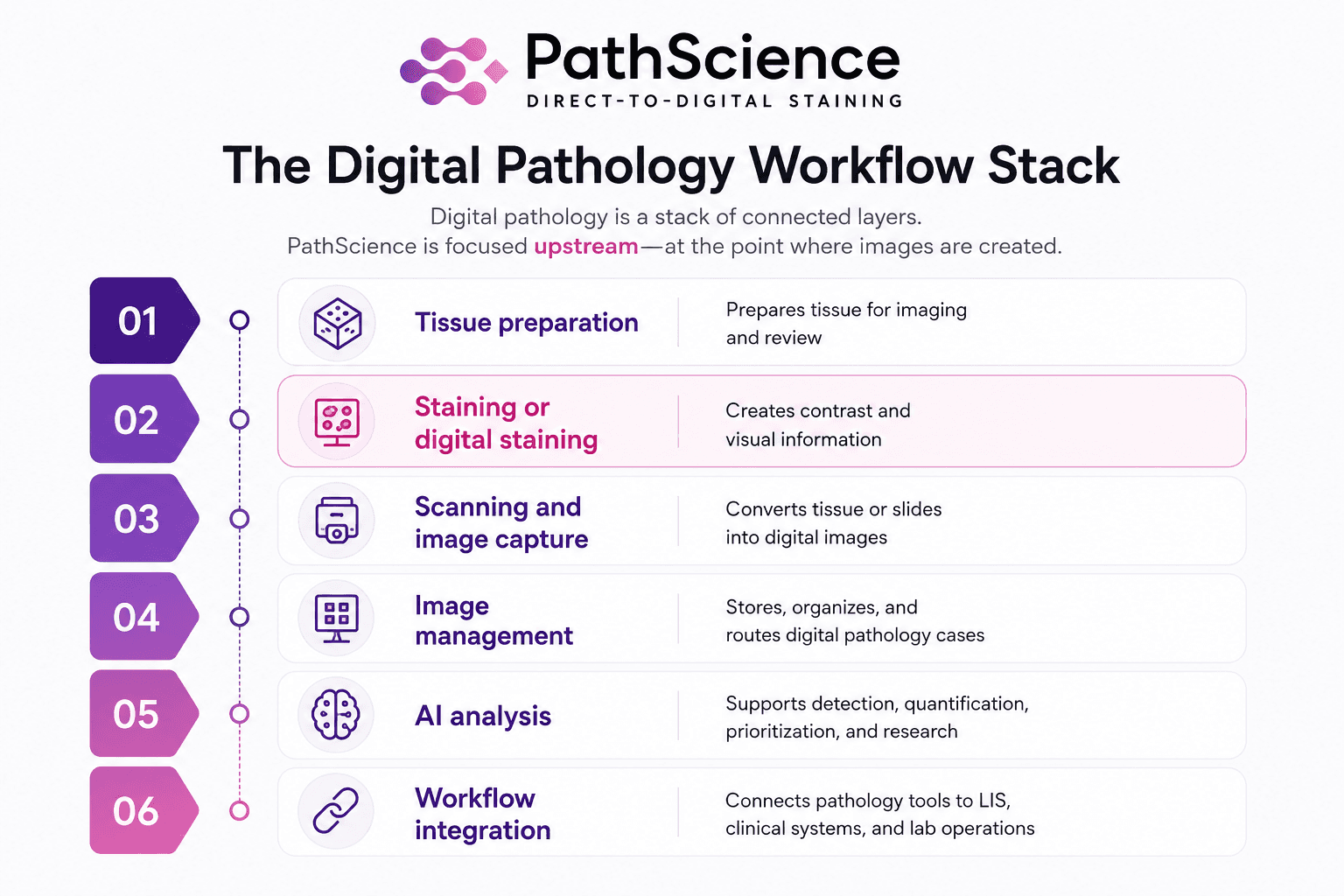
Image management platforms and AI analysis tools are important parts of this stack. But digital staining sits further upstream, closer to the moment when the pathology image is created.
Upstream tools can influence everything from: scan quality, image review, workflow speed, tissue usage, to downstream analysis.
Why this is important for dermatopathology and Mohs surgery
Dermatopathology and Mohs surgey workflows are frequently time-sensitive, visually demanding, and operationally constrained. In Mohs surgery, for example, tissue margin assessment can influence same-day surgical decisions. In dermatopathology, tissue architecture, cellular detail, consistency, and turnaround time all matter.
A digital staining workflow may offer practical advantages in these settings:
It may reduce staining-related waiting time.
It may preserve tissue for additional analysis.
It may reduce dependence on chemical reagents.
It may support more consistent image generation.
It may fit into digital pathology workflows that pathologists are already beginning to use.
Digital staining is not the whole stack, but a very important layer of the stack
Digital pathology includes scanners, viewers, image management systems, AI analysis tools, workflow software, research platforms, clinical validation programs, and laboratory operations.
Digital staining is one layer of that stack.
That means the category does not have to be winner-take-all. A digital staining system still needs scanning, viewing, storage, workflow integration, and pathologist review. A digital pathology platform may eventually benefit from upstream image-generation technologies.
The more useful question is not which layer replaces the others but how the layers work together.
What still has to be proven
Digital staining is promising, but serious pathology tools need serious validation.
For any digital staining workflow to earn trust, several questions matter:
Are the images consistently high quality?
Do pathologists recognize the relevant tissue structures?
How does performance vary by tissue type, preparation method, scanner, and workflow?
How does the digital stain compare with traditional H&E in the intended use case?
What are the limitations?
How should the product be used, and what claims are appropriate?
What regulatory pathway applies?
These questions are not barriers to innovation. They are the work required to make innovation useful.
Pathology is a clinical discipline. New tools have to fit the realities of laboratory operations, pathologist review, regulatory expectations, and patient care.
PathScience’s view
Digital pathology is moving beyond digitization.
The first wave helped pathology images become digital. The next wave will ask how digital tools can improve the way those images are created, reviewed, and integrated into care and research workflows.
At PathScience, we believe Direct-to-Digital Staining can become an important upstream layer in that future.
Our focus is practical: generate familiar H&E-style virtual stain images from unstained tissue scans, support pathologist-centered workflows, and begin with use cases where speed, tissue preservation, and consistency matter.
Digital pathology is no longer just about viewing the slide.
It is about rethinking the workflow around the slide.
